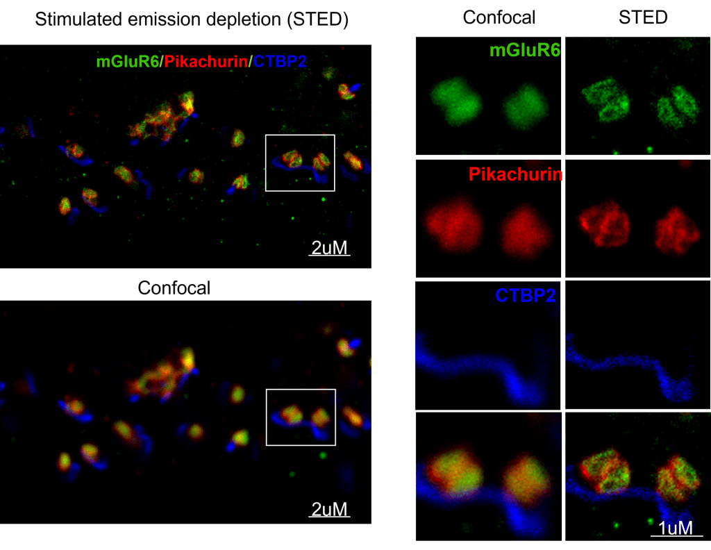
We use both conventional confocal microscopy and super resolution microscopy including STORM and STED to examine the synaptic, subsynaptic and even single molecule scale structural changes.
We use both conventional confocal microscopy and super resolution microscopy including STORM and STED to examine the synaptic, subsynaptic and even single molecule scale structural changes.
