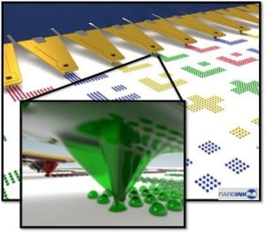
DPN is relatively a new and versatile patterning technique to design, synthesis, and characterization of biomaterials at the nanoscale. This direct-write patterning approach relies on atomic force microscopy (AFM) to transfer material “inks” from the probe tip to a surface with nanometer-scale spatial resolution. DPN facilitates precise patterning of biologically-relevant nanoparticles and molecules for applications in biosensing/imaging, targeted drug delivery, and tissue engineering. DPN will allow control of the size, shape, and chemical functionality of nanoscale biomaterials and these properties are believed to have a profound influence on the ability to direct multi-scale processes including protein function, cellular organization, and tissue regeneration.
In one focused project, we use DPN to better understand scaffold/substrate and scaffold/cell binding in tissue engineering applications. Studies will include deposition of biocompatible polymers (with or without biomolecules) and calcium phosphate-based inks onto a range of substrates in order to understand the binding between cells and surfaces as well as to explore how specific patterns/shapes affect cell morphology and behavior. In addition, multiple patterned hydrogel structures, each with a different cell binding protein or peptide can be investigated. Another focus will be to create fluorescent nanodiamond (ND) color centers as a probe for intracellular tracking, imaging, and drug delivery. Surface functionalization of ND by carboxyl groups leads to high affinity for proteins or other biomolecules either by physical (electrostatic) or chemical (covalent) bonding interactions. Therapeutic molecules conjugated to ND will be investigated for targeted drug delivery.
